Finding the source of chemical reactions
Scientists are constantly searching for the source of things like the origin of the universe, matter or life. Scientists at the U.S. Department of Energy's (DOE) Argonne National Laboratory, in a collaboration with the Massachusetts Institute of Technology (MIT) and several other universities, have demonstrated a way to experimentally detect the most hidden aspect of all chemical reactions - the extremely short-lived transition state that occurs at their initiation. This pivotal discovery could become instrumental in gaining the ability to predict and externally control the outcomes of chemical processes.
"The transition state is key in all of chemistry because it controls the products of molecular reactions," said Kirill Prozument, lead author and chemist in Argonne's Chemical Sciences and Engineering division. Armed with more complete knowledge of certain chemical reactions starting from the transition state, researchers might be able to improve industrial processes involving production of enormous quantities of a chemical -- saving tremendous amounts of energy and money, as well as reducing waste. The same principle might also find application in the synthesis of new, life-saving drugs.
The life of this transition phase is brief, as short as quadrillionths of a second. The problem has been that it has not been possible to experimentally observe the structure of this fleeting state or even to extract sufficient details about it indirectly from the chemical products created by it, until now.
"Physicists cannot directly observe the Big Bang, which happened almost 14 billion years ago, or the transition state that led to the formation of our universe," explained Prozument. "But they can measure various messengers remaining from the Big Bang, such as the current distribution of matter, and thereby uncover many things about the origin and evolution of our universe. A similar principle holds for chemists studying reactions."
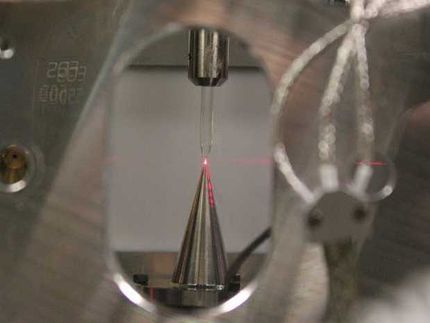
Central to this achievement is the team's experimental technique, chirped-pulse millimeter-wave spectroscopy, which allows characterization of multiple competing transition states on the basis of the vibrationally excited molecules that result in the immediate aftermath of a reaction. This technique is unrivalled in its precision at determining molecular structure and resolving transitions that originate from different vibrational energy levels of the product molecules.
Many hands contributed to the refinement of this experimental technique to expand its scope from the microwave to millimeter region, including Prozument and Robert Field, the Robert T. Haslam and Bradley Dewey Professor of Chemistry at MIT and the senior author on the study.
With this powerful technique, the team analyzed the reaction between vinyl cyanide and ultraviolet light produced by a special laser, which forms various products containing hydrogen, carbon and nitrogen. They were able to measure the vibrational energies associated with the newly formed product molecules and the fractions of molecules in various vibrational levels. The former indicates the amplitudes of which atoms within a molecule move relative to each other. The latter provides information about the geometry of groups of atoms at the transition state as they are giving birth to a product molecule -- in this case, the extent of bending excitation in the bond angle between the hydrogen, carbon and nitrogen atoms. Based upon their measurements, the team identified two transition states that govern different pathways by which the molecule hydrogen cyanide (HCN) springs to life from the reaction.
"Our work demonstrates that the experimental technique works in principle," Prozument says. "The next step will be to apply it to more complex reactions and different molecules." The team's work could thus one day have a major impact on the field of chemistry.
Original publication
Other news from the department science

Get the analytics and lab tech industry in your inbox
By submitting this form you agree that LUMITOS AG will send you the newsletter(s) selected above by email. Your data will not be passed on to third parties. Your data will be stored and processed in accordance with our data protection regulations. LUMITOS may contact you by email for the purpose of advertising or market and opinion surveys. You can revoke your consent at any time without giving reasons to LUMITOS AG, Ernst-Augustin-Str. 2, 12489 Berlin, Germany or by e-mail at revoke@lumitos.com with effect for the future. In addition, each email contains a link to unsubscribe from the corresponding newsletter.


