Super-resolved imaging of a single cold atom on a nanosecond timescale
Scientists have made important progress in the research of cold atom super-resolution imaging
The team of academician GUO Guangcan of University of Science and Technology of China (USTC) of the Chinese Academy of Sciences has made important progress in the research of cold atom super-resolution imaging. The team achieved super-resolution imaging of a single ion in an ion trap system. The results were published in Physical Review Letters.

Symbolic image
Unsplash
The cold atom system is an ideal experimental platform for studying quantum physics, as well as an important physical system for experimental research on quantum simulation, quantum computing, and quantum precision measurement. One of the core experimental techniques in the cold atom system is high-resolution single-particle imaging. In the past ten years, the microscopic imaging technology of the cold atom system has developed rapidly. However, the newly developed technologies are still limited by the fundamental optical diffraction limit, and the resolution can only reach the order of optical wavelength. It is difficult to study quantum phenomena related to the details of the wave function. To study such problems requires optical super-resolution imaging.
Optical super-resolution imaging has developed into a mature tool in the fields of chemistry and biology. However, due to the complexity of cold atom experiments, it is extremely challenging to apply super-resolution imaging technology to cold atom systems. Prior to this, the world has not yet made progress on the direct super-resolution imaging of single atoms (ions).
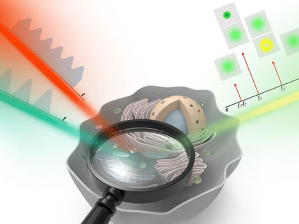
In this study, the researchers adopted the main idea of the Stimulated Emission Depletion (STED) microscopy in the classical super-resolution imaging field, and combining it with the atomic quantum state initialization and reading technology of the cold atom system. They realized super-resolved imaging of a single cold atom (ion) directly for the first time.
Experimental results showed that the spatial resolution of the imaging method can exceed the diffraction limit by more than one order, and the imaging resolution of 175 nm can be achieved by using an objective lens with a numerical aperture of only 0.1.
To further demonstrate the time resolution advantage of this method, the researchers achieved both a time resolution of 50 ns and a single ion positioning accuracy of 10 nm, and used this method to clearly capture the rapid harmonic oscillations of the ion in the trap. Theoretically, by increasing the numerical aperture of the imaging objective and the center extinction ratio of the depleted light (the doughnut spot), the spatial resolution can be further improved to below 10 nm.
This experimental technique can be extended to the multi-body and correlation measurement of cold atom systems, and has good compatibility with other cold atom systems. It can be applied to optical lattices, neutral atom optical tweezers, and cold atom-ion hybrid systems.
Original publication
See the theme worlds for related content
Topic world Fluorescence microscopy
Fluorescence microscopy has revolutionized life sciences, biotechnology and pharmaceuticals. With its ability to visualize specific molecules and structures in cells and tissues through fluorescent markers, it offers unique insights at the molecular and cellular level. With its high sensitivity and resolution, fluorescence microscopy facilitates the understanding of complex biological processes and drives innovation in therapy and diagnostics.


