Overcoming the optical resolution limit
Microsphere assistance enables interferometric topography measurements
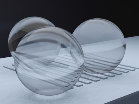
When measuring with light, the lateral extent of the structures that can be resolved by an optical imaging system is fundamentally diffraction limited. Overcoming this limitation is a topic of great interest in recent research, and several approaches have been published in this area. In a recent study published in the Journal of Optical Microsystems, a team of researchers from the University of Kassel in Germany present an approach that uses Microspheres placed directly on the surface of the object to extend the limits of interferometric topography measurements for optical resolution of small structures.

Microspheres placed on a specimen in an interference microscope setup enable topographic measurements of structures below the physical resolution limit.
The Authors
Imaging below the resolution limit is often achieved with systems that use probe labeling, such as fluorescence microscopy, which requires preparation of the sample. Other systems, such as atomic force microscopes, can provide 20 times better lateral resolution than diffraction-limited optical systems. However, they rely on tactile measurement principles that may be unsuitable for certain applications, especially in bio-imaging. Therefore, microsphere assistance can provide a solution for fast and label-free imaging below the diffraction limit.
A Linnik interferometer setup comprising two highly resolving microscope objectives provides fast and non-contact topography measurements of fine structures. Performing a depth scan enables the acquisition of phase information that can be used to reconstruct the surface topography. With an additional microsphere in the imaging path, the physical diffraction limit of this system is extended.
Although experimental studies showed promising results, theoretical explanations considering the relevant imaging mechanisms enabling the improved resolution remained unclear till now. The relevant mechanisms were examined by means of analysis in the 3D spatial frequency domain as well as by comparison with rigorous simulations and ray tracing computations. Investigations in the Fourier domain give the spatial frequencies transmitted by the microsphere into the far field and obtained by the microscope objective. In combination with the rigorous simulations of the resulting near field, this allows a complete simulation of the imaging process with microspheres, and thus, extensive investigations can be performed. In addition, ray tracing enables the investigation of the propagation of individual light rays inside the microsphere and, therefore, contributes to a better understanding of the major physical effects.
"In recent research as well as in industrial applications, there is a need for fast measurement systems below the physical resolution limit that do not require extensive sample preparation. Microsphere-assisted interference microscopy enables such optical topographic surface measurements, and this work contributes to a deeper understanding of the underlying physical mechanisms," said Lucie Hüser, lead author of the paper.
The researchers’ findings provide helpful tools for a deeper understanding of microsphere-assisted interferometry, which can be used to enlarge the knowledge about physical mechanisms in microsphere-assisted interferometry. Furthermore, the effective enlargement of the numerical aperture of the system including the microsphere and the rather small field of view under the microsphere is likely the most relevant mechanism enabling topographical measurements below the resolution limit.
Original publication
See the theme worlds for related content
Topic world Fluorescence microscopy
Fluorescence microscopy has revolutionized life sciences, biotechnology and pharmaceuticals. With its ability to visualize specific molecules and structures in cells and tissues through fluorescent markers, it offers unique insights at the molecular and cellular level. With its high sensitivity and resolution, fluorescence microscopy facilitates the understanding of complex biological processes and drives innovation in therapy and diagnostics.


