Measuring Organ Development
Metrics for organ development have now been defined for the first time
Researchers from Dresden and Vienna reveal link between connectivity of three-dimensional structures in tissues and the emergence of their architecture to help scientists engineer self-organising tissues that mimic human organs.

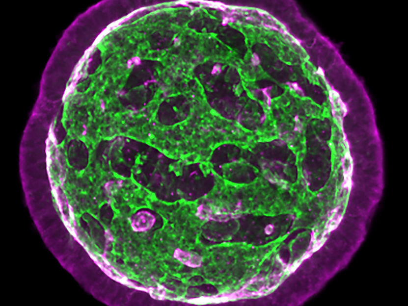
The complex architecture of neuroepithelial organoids emerges from epithelial fusion processes. The cell membranes are marked in red, while the fluid-filled tubes are marked in green.
Keisuke Ishihara et al., Nature Physics (2022)
Organs in the human body have complex networks of fluid-filled tubes and loops. They come in different shapes and their three-dimensional structures are differently connected to each other, depending on the organ. During the development of an embryo, organs develop their shape and tissue architecture out of a simple group of cells. Due to a lack of concepts and tools, it is challenging to understand how shape and the complex tissue network arise during organ development. Metrics for organ development have now been defined for the first time by scientists from the Max Planck Institute of Molecular Cell Biology and Genetics (MPI-CBG) and the MPI for the Physics of Complex Systems (MPI-PKS), both in Dresden, as well as the Research Institute of Molecular Pathology (IMP) in Vienna. In their study, the international team of researchers provide the necessary tools to transform the field of organoids – miniature organs – into an engineering discipline to develop model systems for human development.
The collective interaction of cells leads to the shaping of an organism during development. The different organs feature various geometries and differently connected three-dimensional structures that determine the function of fluid-filled tubes and loops in organs. An example is the branched network architecture of the kidney, which supports the efficient filtration of blood. Observing embryonic development in a living system is hard, which is why there are so few concepts that describe how the networks of fluid-filled tubes and loops develop. While past studies have shown how cell mechanics induce local shape changes during the development of an organism, it is not clear how the connectivity of tissues emerges. By combining imaging and theory, the researcher Keisuke Ishihara started to work on this question first in the group of Jan Brugues at the MPI-CBG and MPI-PKS. He later continued his work in the group of Elly Tanaka at the IMP. Together with his colleague Arghyadip Mukherjee, formerly a researcher in the group of Frank Jülicher at MPI-PKS, and Jan Brugués, Keisuke used organoids derived from mouse embryonic stem cells that form a complex network of epithelia, which line organs and function as a barrier. “I still remember the exciting moment when I found that some organoids had transformed into tissues with multiple buds that looked like a bunch of grapes. Describing the change in the three-dimensional architecture during development proved to be challenging, though,” remembers Keisuke and adds, “I found that this organoid system generates astonishing internal structures with many loops or passages, resembling a toy ball with holes.”
Studying the development of tissues in organoids has several advantages: they can be observed with advanced microscopy methods, making it possible to see dynamic changes deep inside the tissue. They can be generated in large numbers and the environment can be controlled to influence development. The researchers were able to study the shape, number, and connectivity of the epithelium. They tracked the changes in the internal structure of organoids over time. Keisuke continues, “We discovered that tissue connectivity emerges from two different processes: either two separate epithelia fuse or a single epithelium self-fuses by fusing its two ends, and thereby creating a doughnut shaped loop.” The researchers suggest, based on theory of epithelial surfaces, that the inflexibility of epithelia is a key parameter that controls epithelial fusion and in turn the development of tissue connectivity.
The supervisors of the study, Jan Brugues, Frank Jülicher, and Elly Tanaka conclude, “We hope that our findings will lead to a fresh view of complex tissue architectures and the interplay between shape and network connectivity in organ development. Our experimental and analysis framework will help the organoid community to characterise and engineer self-organising tissues that mimic human organs. By revealing how cellular factors influence organ development, these results may also be useful for developmental cell biologists who are interested in organisational principles.”


