Tracking Molecules at Turbo Speed
Researchers devise method to speed up observations of high-throughput microbiological process
Being able to observe micro-organisms and their cellular components is key to understanding fundamental processes that go on inside cells—and thus potentially developing new medical treatments. Microbiologists and biophysicists from the University of Bonn have now developed a method that makes the high-throughput process for observing molecules five times faster, enabling insights to be gained into hitherto unknown cellular functions. The results were published in Nature Methods.
If our skin spends too long exposed to UV rays, e.g. from the sun, it can cause mutations in our DNA, which can potentially lead to cancer. However, the human body has a defense mechanism that it can deploy. “Damage to our DNA activates molecules that repair it quickly, ideally before the cell divides and the damage spreads,” explains Koen Martens from the Institute for Microbiology and Biotechnology at the University of Bonn. Yet nobody quite knows exactly how fast this cellular repair function works, something that Martens now wants to find out.
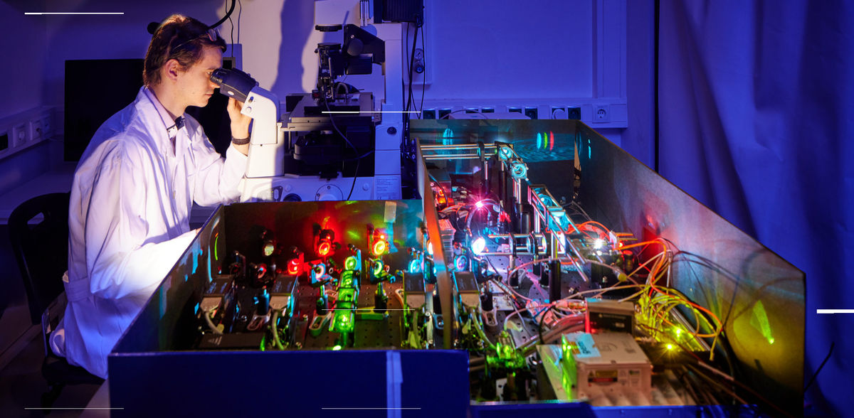
This is easier said than done, however, as the methods used to date are not powerful enough to track individual molecules accurately. “Single particle tracking involves marking the molecule with fluorescent light, making it into a kind of light bulb,” Koen Martens explains. “We then take hundreds of photos a second using a high-resolution microscope. Our ‘light bulb’ lights up the molecule in the darkness of the cell, allowing us to observe it and track its movement over time. This enables us to measure its diffusion and how it interacts with other cellular components.” By looking at the gaps between molecules and the distances traveled by a single molecule from one photograph to another, the researchers can tell whether the particles are moving freely inside the cell or interacting with other molecules. As far as DNA repair is concerned, this indicates when the enzymes are performing their repair work—i.e. when they are interacting with the DNA—and when they are “idle,” i.e. diffusing freely inside the cell.
However, the method does have one drawback: “It’s hard to track multiple molecules at the same time,” Martens explains. “When their paths cross or they’re too close together, you get two light bulbs merging, in effect. Then it’s impossible to identify their movements.” Up until now, therefore, the microbiologists have had to study molecules one after the other in a time-consuming process that is too long-winded to observe the DNA-repairing molecules “at work.” In fact, single particle tracking currently takes longer than the repair process itself.
To solve the problem, Koen Martens has created a piece of software to speed up the high-throughput process. TARDIS (short for “temporal analysis of relative distances”) runs an all-to-all analysis of the distances between locations, i.e. the positions of the molecule in the individual photographs, with increasing time lags. Instead of focusing on individual points as before, it looks at the entire sequence of movements within the cell and thus scrutinizes all the molecules simultaneously. “TARDIS makes the measurement process at least five times faster without any loss of information,” says a happy Martens.
This means that he can now devote his attention to the remaining part of his research project, using TARDIS to study the processes involved in DNA repair in more detail. “I’m especially interested in investigating how easy or difficult certain kinds of damage are to repair and how badly the DNA is damaged by a specific dose of UV radiation or chemicals."
Original publication
Other news from the department science

Get the analytics and lab tech industry in your inbox
By submitting this form you agree that LUMITOS AG will send you the newsletter(s) selected above by email. Your data will not be passed on to third parties. Your data will be stored and processed in accordance with our data protection regulations. LUMITOS may contact you by email for the purpose of advertising or market and opinion surveys. You can revoke your consent at any time without giving reasons to LUMITOS AG, Ernst-Augustin-Str. 2, 12489 Berlin, Germany or by e-mail at revoke@lumitos.com with effect for the future. In addition, each email contains a link to unsubscribe from the corresponding newsletter.
Most read news
More news from our other portals
See the theme worlds for related content
Topic World Cell Analysis
Cell analyse advanced method allows us to explore and understand cells in their many facets. From single cell analysis to flow cytometry and imaging technology, cell analysis provides us with valuable insights into the structure, function and interaction of cells. Whether in medicine, biological research or pharmacology, cell analysis is revolutionizing our understanding of disease, development and treatment options.
Topic world Fluorescence microscopy
Fluorescence microscopy has revolutionized life sciences, biotechnology and pharmaceuticals. With its ability to visualize specific molecules and structures in cells and tissues through fluorescent markers, it offers unique insights at the molecular and cellular level. With its high sensitivity and resolution, fluorescence microscopy facilitates the understanding of complex biological processes and drives innovation in therapy and diagnostics.


