With the Flick of a Switch: Shaping Cells with Light
The therapeutic potential of this research is immense
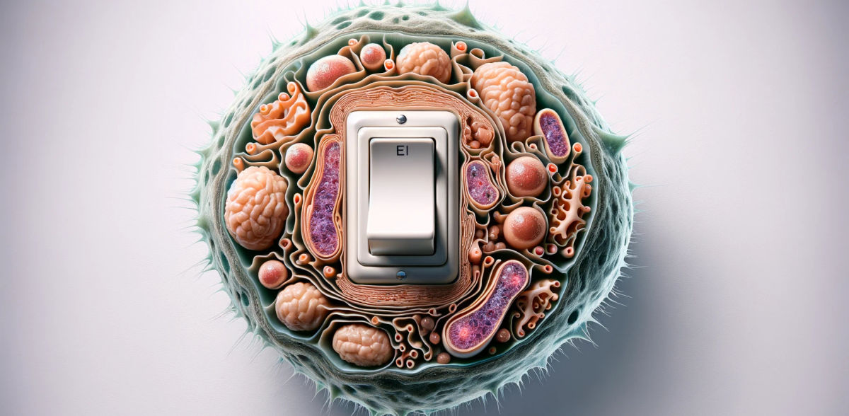
Imagine switching on a light and being able to understand and control the inner dynamics of a cell. This is what the Dimova group has achieved: by shining lights of different colors on replicates of cells, they altered the interactions between cellular elements. Controlling these complex interactions enables us to deliver specific drugs directly into the cells. And with the flick of a switch, we could adjust or even reverse this delivery, potentially revolutionizing the treatment of cells in a smart, accurate and non-invasive way.

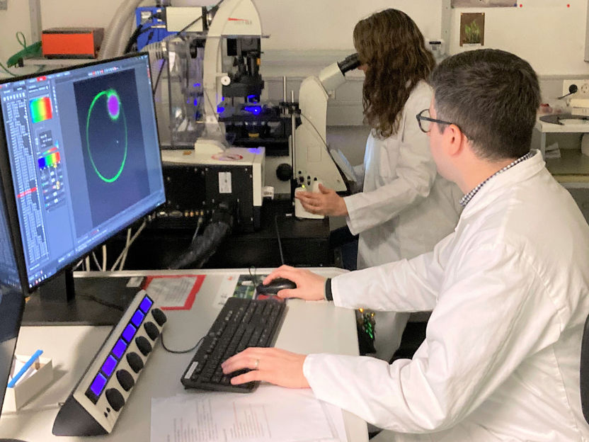
Mina Aleksanyan and Agustin Mangiarotti recording the membrane response at the confocal microscop
© Max Planck Institute of Colloids and Interfaces
Cells are the building blocks of our body and are organized into smaller components, each with a specialized function. Most of these components are enveloped by a protective membrane made of fats. All, with the exception of the biomolecular condensates. These tiny, dynamic droplets prepare the cell for rapid stress response by gathering and organizing repair molecules (among other functions). Rumiana Dimova and her team at the Max Planck Institute of Colloids and Interfaces study the many and intricate ways in which condensates interact with membranes, and how they affect each other’s shape and structure. In their latest work, the researchers have focused on the process of endocytosis. This is how a cell wraps its outer membrane around nutrients or pathogens to ‘eat’ them.
The researchers designed their own lab-made, simplified versions of cells – called giant vesicles – to simulate the cellular processes and analyze them under the microscope. They introduced fats that react to light (“photoswitchable lipids”). Dimova and her team observed how the membranes behaved when exposed to light of different colors: they changed their size and triggered various interactions with the condensates.
“When we shine ultraviolet light on a membrane, it grows and 'swallows’ the condensates,“ – explains Agustín Mangiarotti. “ And we can also reverse the process by switching to blue light,” adds Mina Aleksanyan, “so that the membrane shrinks, expelling the condensate.” This is clearly visible in the video captured using a fluorescence microscope.
The therapeutic potential of this research is immense. The combined use of giant vesicles and light could represent a non-invasive medical treatment to control cellular dynamics.
Here is why. Light is inexpensive and sustainable. And because giant vesicles are synthetic (made in a lab), scientists can use them to probe several dynamics without resorting to culture cells from living organisms. And there is more. Giant vesicles are biomimetic – constructed from molecules found in the human body, such as fats and proteins. They are like tiny capsules that can carry drugs and then fuse organically with cells.
"Now we know that by modulating light we can control how vesicles shape the inner environment of a cell, which could help treat cellular disorders. It's like being able to sculpt a cell from the inside by flicking a light switch," concludes Dimova.
Original publication
Original publication
Agustín Mangiarotti, Mina Aleksanyan, Macarena Siri, Tsu‐Wang Sun, Reinhard Lipowsky, Rumiana Dimova; "Photoswitchable Endocytosis of Biomolecular Condensates in Giant Vesicles"; Advanced Science, 2024-4-6
Organizations
Other news from the department science

Get the analytics and lab tech industry in your inbox
By submitting this form you agree that LUMITOS AG will send you the newsletter(s) selected above by email. Your data will not be passed on to third parties. Your data will be stored and processed in accordance with our data protection regulations. LUMITOS may contact you by email for the purpose of advertising or market and opinion surveys. You can revoke your consent at any time without giving reasons to LUMITOS AG, Ernst-Augustin-Str. 2, 12489 Berlin, Germany or by e-mail at revoke@lumitos.com with effect for the future. In addition, each email contains a link to unsubscribe from the corresponding newsletter.
Most read news
More news from our other portals
Last viewed contents
Tiny laser sensor heightens bomb detection sensitivity

A new animal model to understand metastasis in sarcomas
PerkinElmer Announces Global R&D Expansion in Singapore - New Center of Excellence Focused on Solutions for Environmental Health, Food Safety and Drug Discovery


