Rechargeable nanotorch
Afterglow luminescence imaging tracks cell-based microrobots in real time
An afterglow luminescent nanoprobe opens up new possibilities for imaging living cells. As a research team reports in the journal Angewandte Chemie, their new “nanotorch” can continue to luminesce for over ten days after a single excitation. This allows the routes taken through the body by microrobots to be tracked in real time. In addition, it can be “recharged” non-invasively with near-infrared (NIR) light in a non-contact manner.
© Wiley-VCH
Macrophages are important immune cells that “eat” bacteria as well as being involved in the disposal of cancer cells. In addition, they can take up drugs and transport them into cells, including tumor cells. If they take up magnetic nanoparticles, macrophages can be guided by magnet to a target area within the body, such as a tumor. This allows macrophage “microrobots” to reduce the side effects associated with chemotherapy.
It would be useful to be able to track the microrobots over time as they move through the body. Fluorescence imaging techniques have been considered but require constant external irradiation. This causes is a high level of background noise resulting from the autofluorescence of many biomolecules. In addition, the limited penetration depth of the visible and UV light through tissues required into the tissue limits the depth of detection. One alternative could be the use of probes that can be irradiated before the procedure and produce an afterglow. However, inorganic nanoparticles with long-lasting afterglow harbor the risk that heavy metal ions will leak out; while organic compounds only luminesce for a short time and cannot be repeatedly excited.
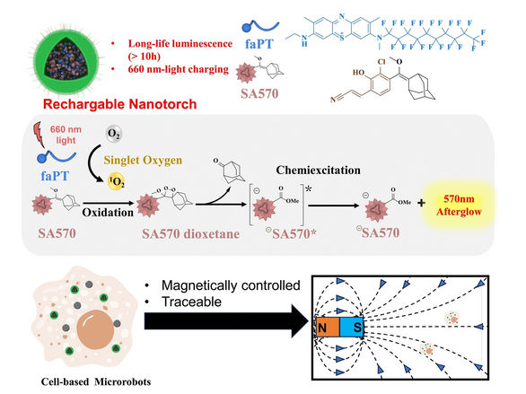
A team from the Shenzhen Institute of Advanced Technology, Chinese Academy of Sciences (China) collaborating with Koç University (Turkey) has now developed a “rechargeable nanotorch”. It is made of multiple components: nanoparticles of a precursor to a luminescent organic molecule, photosensitizers (a hydrophobic analog of methylene blue), and polyethylene glycol equipped with cell-penetrating peptides. The photosensitizer absorbs NIR light and excites surrounding oxygen molecules. This highly reactive singlet oxygen then binds to the precursor and forms a dioxetane group, a four-membered ring made of two oxygen and two carbon atoms. This undergoes a rearrangement that releases the desired luminescent molecule and emits excess energy by luminescing. After the initial irradiation, the nanotorches continue to luminesce for ten days.
Once depleted, the nanotorches can be “remotely” recharged and made to luminesce again by external radiation with NIR light, which can penetrate deep into tissues – multiple times. This requires the relative amounts of photosensitizer and luminescent molecule precursor to be selected so that only some of the precursors are activated with each irradiation. This allows for imaging over longer periods of time.
The China team led by Pengfei Zhang, Ping Gong and Lintao Cai collaborated with the Turkey team with led by Safacan-Kolemen introduced these new nanotorches into macrophage-based microrobots and were able to follow their magnet-guided path through the bodies of mice in real time through the luminescence signals.
Original publication
Most read news
Original publication
Gongcheng Ma, Musa Dirak, Zhongke Liu, Daoyong Jiang, Yue Wang, Chunbai Xiang, Yuding Zhang, Yuan Luo, Ping Gong, Lintao Cai, Safacan Kolemen, Pengfei Zhang; "Rechargeable Afterglow Nanotorches for In Vivo Tracing of Cell‐Based Microrobots"; Angewandte Chemie International Edition, 2024-3-25
Topics
Organizations

Get the analytics and lab tech industry in your inbox
By submitting this form you agree that LUMITOS AG will send you the newsletter(s) selected above by email. Your data will not be passed on to third parties. Your data will be stored and processed in accordance with our data protection regulations. LUMITOS may contact you by email for the purpose of advertising or market and opinion surveys. You can revoke your consent at any time without giving reasons to LUMITOS AG, Ernst-Augustin-Str. 2, 12489 Berlin, Germany or by e-mail at revoke@lumitos.com with effect for the future. In addition, each email contains a link to unsubscribe from the corresponding newsletter.
