Scientists develop cost-efficient medical imaging method
Low-field magnetic resonance imaging combined with hyperpolarization
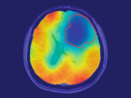
Max Planck scientists presented a low-field magnetic resonance imaging (MRI) scanner for the development of novel MRI methods at the 73rd Meeting of Nobel Laureates in Lindau. As part of an associated scientific event, two researchers from the Max Planck Institute for Biological Cybernetics in Tübingen, Germany, presented a model of a new low-field MRI system. It combines hyperpolarization with imaging techniques that can be run at low magnetic field strengths. The quality of the MRI images can be additionally improved with the help of artificial intelligence.

Max Planck researchers Gabriele Lohmann and Pavel Povolni presented the model of the novel low-field MRI.
Max-Planck-Institut für biologische Kybernetik
Magnetic resonance imaging (MRI) has become the gold standard in clinical diagnostics, especially for the early detection of soft tissue diseases and cancer at an early stage. However, quantitative tumor classification with MRI has so far been difficult due to a lack of high contrast and low sensitivity. The scientists have developed an independent solution in the low-field range of magnetic resonance imaging with a technological process that enables continuous hyperpolarization of the sample itself. Previous hyperpolarization methods could only examine biochemical reactions using a contrast substance injected into the human body. This new method has the potential to expand the existing wide range of applications in magnetic resonance imaging in a cost-effective way and therefore offers the possibility of an affordable diagnostic method for the Global South.
"Our goal is to use our development to contribute to the design of efficient and cost-effective MRI scanners. These can then be optimized to meet the needs of countries in the Global South. That is why we are developing a new, cost-efficient low-field scanner based on second-generation high-temperature superconductors: new polarization processes in combination with deep learning will enable considerably improved imaging than previously known. These will enhance image resolution to such an extent that some medical diagnoses can be made with very high precision," explains project leader Pavel Povolni, who is responsible for the project at the Max Planck Institute for Biological Cybernetics.
The Max Planck Institute for Biological Cybernetics can look back on many years of experience in basic research into medical imaging, in particular magnetic resonance imaging, and is involved in a number of scientific programs dealing with integrative next-generation medical methods. This includes the targeted integration of artificial intelligence. Together with its researchers, the Institute is part of the recently founded Center for Bionic Intelligence in Stuttgart and Tübingen, the ELLIS Society, the Tübingen A.I. Center and the Cluster of Excellence Bionic Intelligence for Health (BI4H), one of six cluster projects at the University of Tübingen.
See the theme worlds for related content
Topic world Diagnostics
Diagnostics is at the heart of modern medicine and forms a crucial interface between research and patient care in the biotech and pharmaceutical industries. It not only enables early detection and monitoring of disease, but also plays a central role in individualized medicine by enabling targeted therapies based on an individual's genetic and molecular signature.


