WITec Honors Scientists with 2024 Paper Awards
The prizes for exceptional scientific publications go to researchers in Great Britain, the Netherlands and South Korea.

Scientists from Great Britain, the Netherlands and South Korea have been recognized with 2024 WITec Paper awards for their work in cellular biology, biomineralization, and single-photon emitting materials, respectively.
The awards highlight three of the many outstanding entries that include data acquired with a WITec microscope. Oxford Instruments WITec congratulates the winners and thanks all the participants for sharing their fascinating work from a wide spectrum of sciences. The jury looks forward to continuing this tradition and receiving many exceptional submissions for the upcoming Paper Award 2025.

The WITec Paper Award Gold winner Dr. Vernon LaLone (left image). Professor Dame Molly Stevens receives the Award from Oxford Instruments representative Stephen McGurk at the University of Oxford (right image) on behalf of her research group, previously of the Imperial College London, UK.
WITec

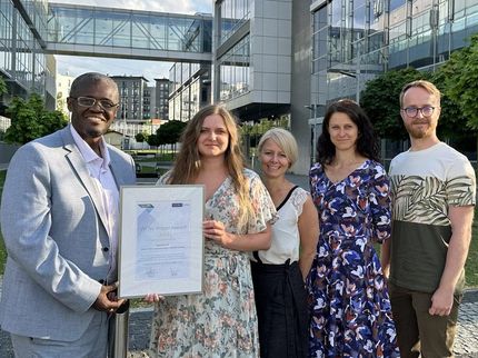
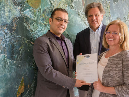
Robin van der Meijden (middle) and his co-authors Prof. Nico Sommerdijk (left) and Dr. Anat Akiva (right) from the Radboud University Medical Center in Nijmegen presenting their Paper Award Silver.
WITec


- GOLD: Vernon LaLone, Aleksandra Aizenshtadt, John Goertz, ..., Steven Ray Wilson, Stefan Krauss, Molly M. Stevens (2023) Quantitative chemometric phenotyping of three-dimensional liver organoids by Raman spectral imaging. Cell Reports Methods 3(4): 100440.
- SILVER: Robin H.M. van der Meijden, Deniz Daviran, Luco Rutten, X. Frank Walboomers, Elena Macías-Sánchez, Nico Sommerdijk, Anat Akiva (2023) A 3D Cell-Free Bone Model Shows Collagen Mineralization is Driven and Controlled by the Matrix. Advanced Functional Materials 33(42): 2212339.
- BRONZE: Michael Neumann, Xu Wei, Luis Morales-Inostroza, Seunghyun Song, Sung-Gyu Lee, Kenji Watanabe, Takashi Taniguchi, Stephan Götzinger, Young Hee Lee (2023) Organic Molecules as Origin of Visible-Range Single Photon Emission from Hexagonal Boron Nitride and Mica. ACS Nano 17(12): 11679-11691.
The Paper Award GOLD: Characterization of biological 3D model systems
Biological and medical research increasingly uses 3D model systems such as organoids to mimic entire organs and study molecular mechanisms in the context of multicellularity in vitro. Vernon LaLone, his colleagues at Imperial College London, UK, and his co-authors at the University of Oslo beautifully presented the potential of Raman imaging for characterizing these complex 3D structures in detail, and in doing so earned this year’s Paper Award GOLD.
The authors developed a method for quantifying the absolute concentration of multiple biological compounds in organoids created from both primary human hepatocytes and human-induced pluripotent stem cells (hiPSCs). Using their approach, even compounds which are generally difficult to visualize with conventional staining techniques were efficiently detected. The newly established quantitative Raman imaging method could identify differences between the primary and stem-cell-derived organoids in terms of their amounts of protein, nucleic acid and unsaturated lipids. In addition, compositional changes were found in the organoids after exposing them to liver-altering drugs. At the same time, the drug’s metabolization and accumulation in the tissue could be tracked.
Together, the authors presented their quantitative Raman method as a powerful tool for the molecular characterization of tissues with high cellular and subcellular resolution. In their words, quantitative Raman imaging “constitutes an important step in developing quantitative label-free interrogation of 3D biological specimens.”
The Paper Award SILVER: Biomineralization of bone structure
Nature is capable of producing an immense variety of materials exhibiting remarkable (mechanical) properties which can only be achieved through controlled formation of the material over multiple length scales. A prime example is bone tissue biomineralization, a process in which minerals are integrated in highly organized organic structures.
This year’s Paper Award SILVER winner Robin H.M van der Meijden and his research group at the Radboud University Medical Center (Radboudumc) in Nijmegen, the Netherlands, studied the biomineralization of bones and determined which components and structures are essential to this process. In their work, they demonstrated the reconstruction of the bone’s main structural component – the osteon – in a cell-free in vitro system. A polarization-resolved Raman microscopy analysis of the generated osteon showed the recapitulation of its chemical and hierarchical 3D structure even in this cell-free environment. In contrast to previous beliefs, not the cellular activity but the structure and composition of the extracellular matrix (ECM) were able to direct the controlled infiltration of minerals into the organic matrix.
In conclusion, the established cell-free in vitro system provided both valuable insight into the processes of bone formation and offers a tool with great potential for future studies on matrix mineralization that “also holds promise for […] mechanistic studies of mineralization related diseases,” as Robin van der Meijden states.
The Paper Award BRONZE: Single-photon emitters in 2D materials
The Paper Award BRONZE winners for 2024, Michael Neumann, Xu Wei and colleagues at the Center for Integrated Nanostructure Physics, Sungkyunkwan University in Suwon, Republic of Korea, working together with partners in Germany and Japan, dove deep into a topic of quantum optics. The subject of their studies was hexagonal boron nitride (hBN), an intensely studied layered material that often features single-photon emitters (SPEs), which are of great interest for quantum technologies. These SPEs had so far been attributed to luminescent point defects in the hBN crystal lattice, however, in their work, the authors questioned this assignment.
By studying the photoluminescent activity of hBN samples they demonstrated that many of the observed SPEs were not hosted by hBN but were in fact fluorescent molecules which resulted from organic processing residues, such as from organic solvents, being trapped at the interface between hBN and its substrate. Supporting this, hBN samples free from organic contamination did not contain SPEs. Moreover, the same class of SPEs could also be formed under sheets of fluorphlogopite mica, a different layered insulator.
Concerning the new understanding of the origin of many of the SPEs observed in hBN, Michael Neumann and Xu Wei commented, “[T]he change of perspective introduced by our work will accelerate the development of hBN-based quantum photonic technologies, by guiding a shift from uncontrolled emitter generation toward rational selection of fluorophore species and their controlled placement, as demanded by many applications in emerging quantum technologies.”
Original publication
Vernon LaLone, Aleksandra Aizenshtadt, John Goertz, Frøydis Sved Skottvoll, Marco Barbero Mota, Junji You, Xiaoyu Zhao, Henriette Engen Berg, Justyna Stokowiec, Minzhi Yu, Anna Schwendeman, Hanne Scholz, Steven Ray Wilson, Stefan Krauss, Molly M. Stevens; "Quantitative chemometric phenotyping of three-dimensional liver organoids by Raman spectral imaging"; Cell Reports Methods, Volume 3
Robin H.M. van der Meijden, Deniz Daviran, Luco Rutten, X. Frank Walboomers, Elena Macías‐Sánchez, Nico Sommerdijk, Anat Akiva; "A 3D Cell‐Free Bone Model Shows Collagen Mineralization is Driven and Controlled by the Matrix"; Advanced Functional Materials, Volume 33, 2023-6-20
Michael Neumann, Xu Wei, Luis Morales-Inostroza, Seunghyun Song, Sung-Gyu Lee, Kenji Watanabe, Takashi Taniguchi, Stephan Götzinger, Young Hee Lee; "Organic Molecules as Origin of Visible-Range Single Photon Emission from Hexagonal Boron Nitride and Mica"; ACS Nano, Volume 17, 2023-6-5
Other news from the department science