Physicist detects movement of macromolecules engineered into our food
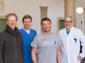
Prof. Rikard Blunck receives the Society of General Physiologists’ Cranefield Award for his breakthrough
Toxin proteins are genetically engineered into our food because they kill insects by perforating body cell walls, and Professor Rikard Blunck of the University of Montreal’s Group for the study of membrane proteins (GÉPROM) has detected the molecular mechanism involved. In recognition of his breakthrough, he received the Traditional Paul F. Cranefield Award of the Society of General Physiologists.
“This study is about gaining a better understanding of the basic functioning of the toxin proteins in order to judge the risks of using them as pesticides for our nutrition,” Dr. Blunck explained.
The Cry1Aa toxin of B. thuringiensis that was investigated is a member of the class of proteins which are called “pore-forming toxins” because they perforate the walls, or membranes, of cells. Cry toxins kill insect larvae if ingested by them and are, therefore, genetically engineered into a number of transgenic crops, including those for human consumption, to make them resistant against these insects.
The pores in the membranes cause minerals necessary for the cell to live to break out and collapse the energy household of the cell. While these toxins could be studied outside of cell membranes through existing techniques that provide images of the 3D structure, the toxins rapidly change their architecture once in contact with the membrane, where the traditional approaches cannot be applied.
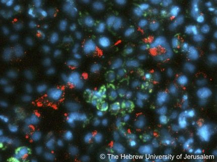
Dr. Blunck and his co-workers found a way of using fluorescent light to analyze the architecture and mechanism of the proteins in an artificial cell wall environment. Planar lipid bilayer (PLB) are artificial 0.1 mm-wide systems that mimic the cell membrane. The researchers developed a chip to investigate proteins introduced into these artificial cell walls with fluorescent light waves. Molecular fluorescent probes are coupled to the toxin proteins. If the proteins now enter the artificial membranes and change their structure, their architecture and movement and even their distribution can be followed – thanks to the developed technique - by the fluorescent light they are emitting.
“By watching the toxin in both its active and inactive state, and by measuring the dynamic changes of the light emitted by the molecular probes, we were able to determine which parts of it were interacting with the membrane to cause the pores.” Dr. Blunck explained. “We expect the technique to be applied to a wide range of disease-causing toxins in future.”
Original publication
Rikard Bunck, Nicolas Groulx and Marc Juteau; "Rapid topology probing using fluorescence spectroscopy in planar lipid bilayer: the pore-forming mechanism of the toxin Cry1Aa of Bacillus thuringiensis”; Journal of General Physiology.
Most read news
Original publication
Rikard Bunck, Nicolas Groulx and Marc Juteau; "Rapid topology probing using fluorescence spectroscopy in planar lipid bilayer: the pore-forming mechanism of the toxin Cry1Aa of Bacillus thuringiensis”; Journal of General Physiology.
Organizations

Get the analytics and lab tech industry in your inbox
By submitting this form you agree that LUMITOS AG will send you the newsletter(s) selected above by email. Your data will not be passed on to third parties. Your data will be stored and processed in accordance with our data protection regulations. LUMITOS may contact you by email for the purpose of advertising or market and opinion surveys. You can revoke your consent at any time without giving reasons to LUMITOS AG, Ernst-Augustin-Str. 2, 12489 Berlin, Germany or by e-mail at revoke@lumitos.com with effect for the future. In addition, each email contains a link to unsubscribe from the corresponding newsletter.
