Nanomedicines on their way through the body
Which pathways do nanomedicines take after they have been swallowed?
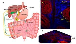
Advances in pharmaceutical nanotechnology have yielded ever increasingly sophisticated nanoparticles for medicine delivery. When administered via oral, intravenous, ocular and transcutaneous delivery routes, these nanoparticles can elicit enhanced drug performance. One such recently developed nanoparticle is Quaternary Ammonium Palmitoyl Glycol Chitosan (GCPQ), a chitosan-based polymeric micelle which can be used to encapsulate drugs and enhance their oral absorption and their intravenous activity by up to one order of magnitude. In spite of its great potential, the mechanisms by which GCPQ micelles – or other nanoparticle-based delivery systems – interact with organs at the cellular scale are not yet clear. However, full knowledge of these mechanisms is a prerequisite for a rational design optimizing their performance.

Natalie Laura Garrett an a team of scientist from the University of Exeter and the UCL School of Pharmacy in London (UK) used multimodal nonlinear optical microscopy to investigate these mechanisms using deuterated GCPQ delivered orally to mice.
They combined coherent anti-Stokes Raman scattering (CARS) microscopy, second harmonic generation (SHG) and two photon fluorescence (TPF) microscopy as a multi-modal label-free method. CARS microscopy has many advantages over conventional imaging including: up to several hundred micron depth penetration into biological tissue; intrinsic optical sectioning and high spatial resolution; label-free chemically specific contrast. When combined with CARS microscopy, TPF and SHG allow detailed three-dimensional visualisation of nanoparticles pinpointed with sub-cellular precision against a complex biological background.
The multi-modal method was used to image three of the most important organs for oral drug delivery: the liver, the intestine and the gall bladder. By doing so, they demonstrated for the first time that orally administered chitosan nanoparticles follow a recirculation pathway from the gastrointestinal tract via enterocytes in the villi, pass into the blood stream and are transported to the hepatocytes and hepatocellular spaces of the liver and then to the gall bladder, before being re-released into the gut together with bile. Such recirculation may also improve drug absorption.
Original publication
N.L. Garret et al.; J. Biophotonics 5, 458-568 (2012).
See the theme worlds for related content
Topic world Fluorescence microscopy
Fluorescence microscopy has revolutionized life sciences, biotechnology and pharmaceuticals. With its ability to visualize specific molecules and structures in cells and tissues through fluorescent markers, it offers unique insights at the molecular and cellular level. With its high sensitivity and resolution, fluorescence microscopy facilitates the understanding of complex biological processes and drives innovation in therapy and diagnostics.


