Benefits of Deep Learning and AI in Microscopy: Label-Free Nucleus Detection in Micro Well Plates
EVIDENT Europe GmbH

New AI approaches have potential to reduce or replace fluorescence markers in live cell imaging
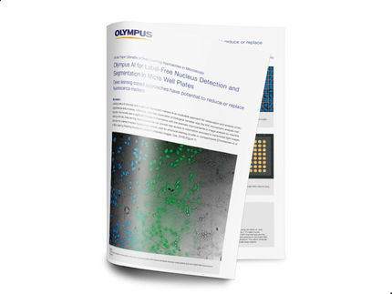
Label-free observation of biological samples has recently seen a significant increase in importance with the dramatic improvements in image analysis by machine learning methods. Deep learning-based approaches can provide new access to information encoded in transmitted light images:
- They have the potential to replace fluorescence markers used for structural staining of cells or compartments.
- They give clear advantages in solving challenges of transmitted light analysis like low contrast, dust, imperfections, particles, condensation artefacts etc.
The white paper explains how Deep Learning Technology, neural networks and the “ground truth” open the door to label-free analysis applications. Based on a typical use case of a whole 96 well plate with variation in buffer filling level, condensation effect, meniscus-induced imaging artefacts, etc. the application is described from the “Training Phase to get the ground truth for label-free segmentation” to detection and validation of results. No manual annotations are required, thanks to the self-learning microscopy approach. Fully automated training data generation enables robust segmentation of nuclei with better accuracy than measurements based on fluorescence.
Use of Olympus’ AI-based approach brings significant benefits to many live-cell analysis workflows. Aside from the improved accuracy, using bright field images also avoids the need for using genetic modifications or nucleus markers. Not only does this save time on sample preparation, it also saves the fluorescence channel for other markers. Furthermore, the shorter exposure times for bright field imaging mean reduced phototoxicity and further time savings on imaging.
Download white paper now

Benefits of Deep Learning and AI in Microscopy: Label-Free Nucleus Detection in Micro Well Plates
New AI approaches have potential to reduce or replace fluorescence markers in live cell imaging