Chasing tiny vehicles
Microscope shows how nanoferries invade cells
nanoparticles are just billionths of a millimeter in size. Exhibiting novel and often surprising properties, they are finding their way into an endless stream of equally innovative products. In medical therapies, for example, tiny nanovehicles could one day ferry drugs or even genes into cells. So far, the only way of testing these approaches has been to wait for the desired effect to show – the activation of a transported gene inside a cell for example. Under the direction of LMU Munich physicochemist Professor Christoph Bräuchle, a research group cooperating with Dr. Christian Plank of the Technische Universität München (TUM) has now used a highly sensitive microscopic technique to pursue individual nanoparticles as they make their way into target cells – in real-time and at high spatial and temporal resolution. They tested magnetic nanoparticles that could be used, among other things, in cancer therapy. This approach should also allow a better understanding of existing nanovectors as well as the development of new systems, as reported in the current cover story of the Journal of Controlled Release.
Nanoparticles are so small that many barriers in the body simply can't stop them. They can also use the bloodstream to reach any part of the body. Researchers and doctors alike hope that these tiny vehicles will one day be put to work in therapies carrying drugs directly to the seat of a disease. "Even genes can be transported this way," says Plank. "That means we could be seeing new breakthroughs in gene therapy soon, which has seen more than its fair share of setbacks. After all, lacking most are functional transporters." Such vehicles or vectors have been developed mainly from viruses until now. But even deactivated viruses can sometimes trigger unwanted side-effects. Nanoferries, on the other hand, have been tailored to deliver genes or drugs directly to the target without side-effects.
For such a targeted delivery, however, nanoferries need a kind of search mechanism to guide them to where their cargo is needed. Magnetic particles have already been tried in cancer therapies: They have been administered by infusion and then directed – via magnetic fields – to a tumor whose cells they should invade directly. But until now, is has been impossible to observe nanoparticles along their route, especially into living tumor cells. It is a prerequisite, though, for therapeutic approval and the definition of functional doses to know the exact path of these carriers and the efficiency of their transport and uptake by cancer cells.
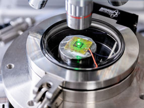
So far, only the appearance or absence of the desired therapeutic effect would tell whether an approach was even promising or not. "It's like a black box," Bräuchle says. "You put something in at one end, then wait and see if anything comes out at the other end. What happens in between is anyone's guess." Now, his workgroup has employed highly sensitive single-molecule fluorescence microscopy to follow the nanoferries on their voyage. This highly sensitive method works by tagging individual particles with a dye that acts like a "molecular lamp" to light up the particle's path into the cell.
"Thus, we have traced magnetic lipoplex nanoparticles and made movies of their transport," reports Anna Sauer, first author of the study. "We were able to watch the particles in real-time and at high temporal and spatial resolution as they made their way into the cells." In doing so, the research team could even define separate phases: how the particles reached the cell membrane, came to rest there and then ultimately – enclosed in a membrane vesicle – invaded the cells. The vesicles move randomly, often downright erratically inside the cell, until a so-called motor protein binds them and quickly transports them towards the cell nucleus – the ultimate target for the gene.
The research team is now in a position to characterize and describe in great detail the individual steps along this path. "Our new approach has also revealed bottlenecks in nanoferry transport," Bräuchle reports. "We saw, for example, that the magnetic field can only direct particles outside cells. But, contrary to expectations, it did not facilitate entry into cells. Thanks to these new insights, existing nanoferries can be suitably optimized in future, and even new systems developed."
Original publication: A.M. Sauer et al.; "Dynamics of magnetic lipoplexes studied by single particle tracking in living cells"; Journal of Controlled Release, 20 July 2009
See the theme worlds for related content
Topic world Fluorescence microscopy
Fluorescence microscopy has revolutionized life sciences, biotechnology and pharmaceuticals. With its ability to visualize specific molecules and structures in cells and tissues through fluorescent markers, it offers unique insights at the molecular and cellular level. With its high sensitivity and resolution, fluorescence microscopy facilitates the understanding of complex biological processes and drives innovation in therapy and diagnostics.
