Rapid diagnosis of diseases with novel blood test
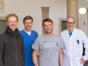
Prof. Dr. Jochen Guck, research group leader at the Biotechnology Center of TU Dresden (BIOTEC), together with medical colleagues from the University Hospital Carl Gustav Carus Dresden and partnering institutes from Dresden (Germany), Cambridge (UK), Glasgow (UK), and Stockholm (Sweden) use a technique called “real-time deformability cytometry” to screen thousands of cells in a drop of blood for unusual appearance and deformability in a matter of minutes. This novel blood test promises to speed up the correct diagnosis of many disease conditions including leukaemia, malaria, bacterial or viral infections, which in turn can lead to a faster and more accurate start of therapy.
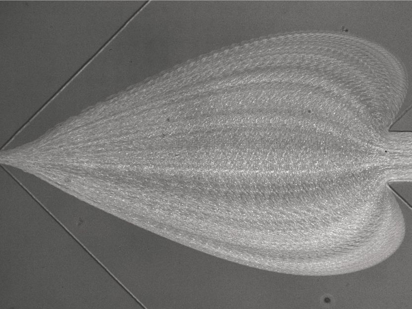
RT-DC in action. The artistic rendering of the microscopic view into the measurement chip shows the trajectories of many individual blood cells flowing from right to left. When encountering sheath flows from top and bottom, they widen to form a “heart” before entering the narrow measurement channel on the left, where the appearance and deformation of the cells are being analysed.
©Daniel Klaue/ZELLMECHANIK DRESDEN GmbH
When you are sick and go to the doctor, it is often not obvious what exactly is wrong — what is causing fever, nausea, shortness of breath or other symptoms. It is important to find this out quickly so that the right action can be taken. One of the first steps is to obtain a blood sample and to count how many of the different blood cells are present in it. This is called a complete blood count, and the information it provides has turned out to be surprisingly useful. A large number of certain white blood cells, for example, can show that the body is fighting an infection. But there might be several reasons why the number of white blood cells has increased, so this information alone is often not enough for a specific diagnosis.
There are many hundreds of possible tests that can supplement the results of a complete blood count. These might identify bacteria or measure the concentrations of certain molecules in the blood, for example. But which test will give the important clue that reveals the source of the illness? This can be difficult to predict. Although each test helps to narrow down the final diagnosis they become increasingly expensive and time-consuming to perform, and rapid action is often important when treating a disease.
Can we get more decisive information from the initial blood test by measuring other properties of the blood cells? The research team now show that this is possible using a technique called “real-time deformability cytometry” (RT-DC). This method forces the blood cells in a small drop of blood to flow extremely rapidly through a narrow microfluidic channel while they are imaged by a fast camera. A computer algorithm can then analyze the size and stiffness of the blood cells in real-time. The research team show that this approach can detect characteristic changes that affect blood cells as a result of malaria, spherocytosis, bacterial and viral infections, and leukemia. Furthermore, many thousands of blood cells can be measured in a few minutes — fast enough to be suitable as a diagnostic test.
„The 36,000x increase in measurement throughput from 100 cells/hour with previous techniques to measure cell stiffness to now 1,000 cells/sec with RT-DC, which we have accomplished in the last few years, was already remarkable. But to now see RT-DC actually being applied to real-world problems and to improve the diagnosis of many diseases, is really gratifying. This is the apex of a research vision I have been following for almost 20 years”, explains Jochen Guck.
These proof-of-concept findings can now be used to develop actual diagnostic tests for a wide range of blood-related diseases. The approach could also be used to test which of several drugs is working to treat a certain medical condition, and to monitor whether the treatment is progressing as planned.
This research was supported by an ERC Starting Investigator Grant “LightTouch”, an Alexander von Humboldt Professorship, the FP7 Marie-Curie Initial Training Network LAPASO, as well as funding from the Saxon Ministry of Science and Art (SMWK) and the European Fund for Regional Development (EFRE). The technology is now being developed into an actual medical device by the TUD spin-off company Zellmechanik Dresden GmbH, supported by the two ERC Proof-of-Concept Grants FastTouch and BASIC.
Original publication

Get the analytics and lab tech industry in your inbox
By submitting this form you agree that LUMITOS AG will send you the newsletter(s) selected above by email. Your data will not be passed on to third parties. Your data will be stored and processed in accordance with our data protection regulations. LUMITOS may contact you by email for the purpose of advertising or market and opinion surveys. You can revoke your consent at any time without giving reasons to LUMITOS AG, Ernst-Augustin-Str. 2, 12489 Berlin, Germany or by e-mail at revoke@lumitos.com with effect for the future. In addition, each email contains a link to unsubscribe from the corresponding newsletter.
More news from our other portals
See the theme worlds for related content
Topic World Cell Analysis
Cell analyse advanced method allows us to explore and understand cells in their many facets. From single cell analysis to flow cytometry and imaging technology, cell analysis provides us with valuable insights into the structure, function and interaction of cells. Whether in medicine, biological research or pharmacology, cell analysis is revolutionizing our understanding of disease, development and treatment options.
Topic world Diagnostics
Diagnostics is at the heart of modern medicine and forms a crucial interface between research and patient care in the biotech and pharmaceutical industries. It not only enables early detection and monitoring of disease, but also plays a central role in individualized medicine by enabling targeted therapies based on an individual's genetic and molecular signature.
