New Opportunity for Cancer Therapy: Miniature Lab Provides Insights into Metastases Development
Fraunhofer IWS Develops Microphysiological Systems for Cultivation of Tumor Tissue Sections in Joint Effort
Every year, around half a million people in Germany are diagnosed with cancer. Despite the existence of effective treatment options for many types of cancer, many questions about the development of the disease remain unanswered. Why does a tumor develop? What factors promote the growth of cancer cells? Why do metastases spread to other organs over time? The animal models mainly in use to date only represent the actual processes in the human body to a limited extent. The Fraunhofer Institute for Material and Beam Technology IWS in Dresden has developed special microsystems in collaboration with the Fraunhofer Institute for Toxicology and Experimental Medicine ITEM in Hanover and the University of Regensburg. They are now using them to examine tissue sections of tumors under realistic conditions.
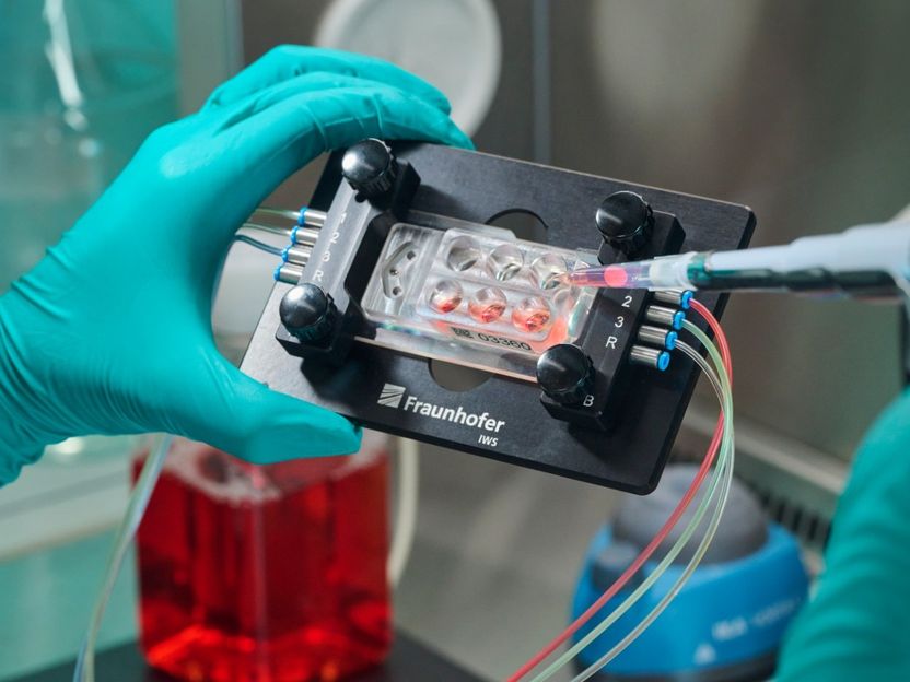
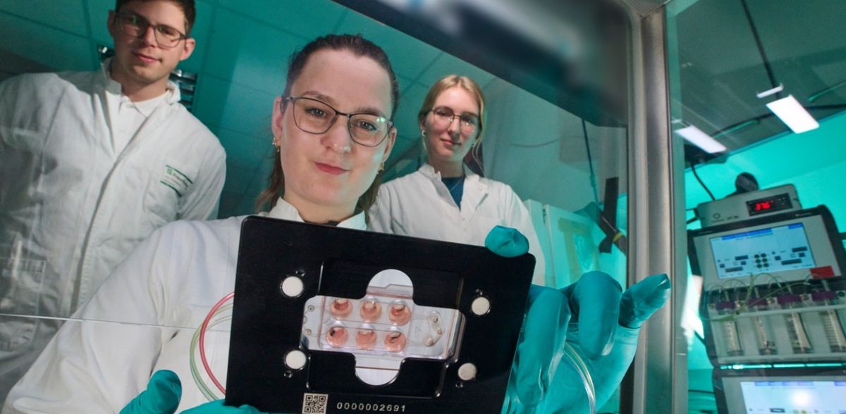
Fraunhofer IWS has been developing microphysiological systems the size of a box of tablets for several years. Cultivating up to ten tissue slices on one chip is now possible.
© minkus-images.de/Fraunhofer IWS
Fraunhofer IWS has been successfully developing microphysiological systems in the size of a tablet box for several years. They can be used to mimic organ function or disease processes in cell culture, to study diseases outside the organism, meaning ex vivo, and to test drugs. “We stack multiple layers of plastic film,” explains Stephan Behrens, development engineer at Fraunhofer IWS. These are structured earlier using lasers to create channels and chambers as well as pumps and valves. They represent different processes of the human body. A blood-like fluid circulates in the microphysiological systems, supplying the cells with oxygen and nutrients. A new challenge in an interdisciplinary project was to investigate the metastasis of tumors in microphysiological systems.
Prof. Christoph Klein, senior professor of Experimental Medicine and Therapy Research at the University of Regensburg and division director of Personalized Tumor Therapy at Fraunhofer ITEM, approached Fraunhofer IWS with this request. Together with the University of Erlangen-Nuremberg, the German Research Foundation had approved a new collaborative research center for the Regensburg researchers in 2020. It aims to uncover how exactly metastases colonize the organs.
Tumor and Immune System Interact on a Chip
“To study this, it was important for us to integrate several tumor tissue sections into our microphysiological system,” says Florian Schmieder, group leader at Fraunhofer IWS. This was achieved for the first time in the world in this project. Up to ten tissue sections can now be cultivated in parallel on one chip. The team at Fraunhofer IWS additionally realized ports from which they can take samples for examination at any time. “We can also continuously measure important parameters such as the CO2 content, pH value, and oxygen concentration,” he continues. “We use these sensors to directly measure directly inside the microphysiological system and they can be reused for further investigations.”
Fraunhofer ITEM experts contributed their knowledge of tissue slices. They opted for ultra-fine sections of lung tissue, explains Prof. Armin Braun, Head of Preclinical Pharmacology and Toxicology at Fraunhofer ITEM. “When operating on a patient with a lung tumor, not only the tumor itself is removed, but also healthy tissue.” A vibratome equipped with an oscillating razor blade produces wafer-thin slices with a thickness of 350 µm and a diameter of around one centimeter from these samples. These are still well supplied with nutrients. When applied to the chip, the tissue slices remain vital and functional in the microphysiological system for an extended period of time. “We can, therefore, observe the interaction of the human immune system with the tumor,” adds Braun. All relevant immune cells are already present in the section. “This means we are very close to the real system, much closer than would be possible with animal models.”
Using Systems to Research Other Diseases
How does cancer develop, and how does it spread in the body? An important point here is that the metabolism in the tumor differs from that in normal tissue. “It is important for clinicians to be able to investigate which conditions in an organ attract metastases,” explains Florian Schmieder. High oxygen concentrations and pH values are crucial for this. The researchers at the Fraunhofer IWS want to adjust these environmental conditions even more effectively in the microsystems in the future. “So far, for example, we have been able to change the oxygen content in the entire system," he continues. One challenge now is to enable different oxygen concentrations on a chip to observe the reaction of the tumor cells and metastases.
Ideally, combining several types of tissue from one patient would be feasible. “Such samples prove scarce in reality,” explains Schmieder. However, combining blood samples and tissue from the same patient in the system is possible. In combination with the various sensors, this results in an added value previously not achievable using other methods. The technology can also serve as a sensible alternative to previous animal experiments. Unfortunately, research cannot yet eliminate the need for animal models.
In parallel, the 15-member team at Fraunhofer IWS is currently working on projects to test the use of tissue slices for other diseases. One example is fibrosis. In this case, the immune system reacts differently to the tissue, which then hardens pathologically and partially loses its function. These processes restrict the function of tissues and organs. “We are working on this issue in the Fraunhofer internal project FIBROPATHS,” says Schmieder. The aim is to clarify which specific systems the individual tissues need in the mini-laboratory to cultivate them for a longer time period.
New Therapies for Cancer Patients Possible
Prof. Christoph Klein is encouraged by the gained research results into tumor growth and metastasis formation with the help of microphysiological systems. “If we want to investigate diseases, this is an interesting new opportunity for us,” says the physician. “Understanding metastasis comprehensively is a key to new therapeutic methods that prevent the subsequent formation of metastases in the bodies of cancer patients.”
Florian Schmieder sees excellent potential for the future in technology: “We are becoming increasingly modular with our systems.” In the future, different components could be combined in new ways to clarify various scientific issues.
