How cells export and embed proteins in the membrane
EMBL scientists first to visualise crucial step
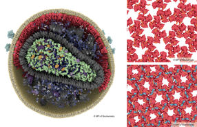
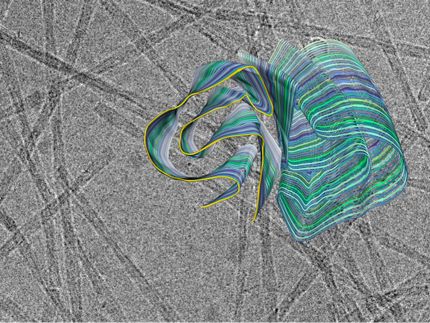
Like an overprotective parent on the first day of school, a targeting factor sometimes needs a little push to let go of its cargo. Scientists at the European Molecular Biology Laboratory (EMBL) in Grenoble, France, have visualised one such hand-over. They were the first to determine the structure of a ribosome-protein complex involved in carrying nascent proteins out of the cell. Their work, published in Nature Structural and Molecular Biology, could increase understanding of illnesses such as cystic fibrosis and some forms of Parkinson's disease, in which improper protein targeting leads proteins to harmfully accumulate inside cells. In most organisms, proteins destined to cross or be embedded in a membrane contain a polypeptide sequence that is recognized during translation by a targeting factor known as the signal recognition particle (SRP). SRP binds to the ribosome synthesizing the polypeptide, and subsequently also binds an SRP receptor, located next to the machinery that transfers proteins across the membrane and out of the cell. EMBL scientists have now generated the first-ever structural image of this important step in the process. "The SRP receptor acts as a switch between the cargo binding and the release," says Christiane Schaffitzel, who led the research at EMBL, "Now we have seen for the first time how the release can happen at a molecular level." Schaffitzel's group is taking structural snapshots of entire pathways by which proteins are synthesized and targeted to their final positions. To capture this hand-over step, the scientists had to overcome the fact that the link between SRP and its receptor is usually transient, chemically unstable. They engineered the SRP receptor so that it would bind more stably to SRP, then introduced ribosomes and observed the resulting complexes using cryo-electron microscopy (cryo-EM). Cryo-EM can be performed in roughly physiological conditions, providing a picture that closely resembles what happens in living cells. This picture can then be combined with higher-resolution crystallography data and biochemical studies – an exciting hybrid approach the EMBL scientists will further exploit to follow protein targeting all the way from start to finish.
Other news from the department science