New tool for cell research may help unravel secrets of disease
Advancements in understanding rotational motion in living cells may help researchers shed light on the causes of deadly diseases, such as Alzheimer's, according to Ning Fang, an associate scientist at the U.S. Department of Energy's Ames Laboratory and faculty member at Iowa State University.
In an article entitled "Resolving Rotational Motions of Nano-objects in Engineered Environments and Live Cells with Gold nanorods and Differential Interference Contrast Microscopy" published in the Journal of the American Chemical Society, and an article in press in ACS Nano, Fang and his research team write about the influence of differential interference contrast microscopy on revealing nanoparticle movement in living cells.
In the human body, numerous biological nanomachines perform various functions. But according to Fang, scientists have only a limited understanding of how these nanomachines work, especially in cellular environments. And because the malfunction of any of these nanomachines can lead to diseases, such as Alzheimer's, there is a great need for new techniques to help investigate the composition, dynamics and working mechanisms of these nanomachines.
To understand how these nanomachines work, scientists look at various types of motion in nanomachines that are essential to their function. Translational motion, or movement in which the position of an object is changed, can be tracked through a variety of current techniques. However, rotational motion, which is as important and fundamental as translational motion, was largely unknown due to technical limitations.
Previous techniques, such as particle-tracking or single-molecule fluorescence polarization, only allowed rotational movement to be resolved in vitro, such as in a Petri dish. In their research, Fang's group has gone beyond studying motions in the in vitro environment to imaging rotational movement in the in vivo, or live cell, environment.
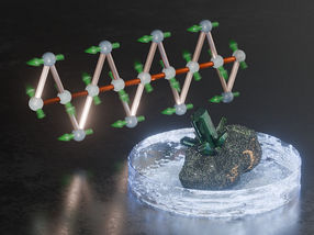
To do this, they rely upon the use of gold nanorods, which are only 25 by 73 nanometers in size (a well-packed bundle of 1000 nanorods has the same diameter as a human hair). In live cells, these non-toxic nanorods scatter light differently depending upon their orientation. Using a technique called differential interference contrast microscopy, or DIC, Fang's team can capture both the orientation and the position of the gold nanorods in addition to the optical image of the cell and, thus, reveal a particle's 5D (3 spatial coordinates and 2 orientation angles) movement within living cells.
Other news from the department science