A well-matched couple
A new setup for hybrid photoacoustic and ultrasound imaging with optical ultrasound detection provides perfectly co-registered dual-modality section images
Photoacoustic (PA) imaging is a method to visualize structures with optical contrast in biological tissue. Despite the strong optical scattering in tissue, high resolution images in the visible and near infrared spectral range can be obtained. The method is based on sound waves which are generated in regions of enhanced optical absorption. The absorption of diffusely propagating optical radiation leads to a temperature rise and thermoelastic expansion causing sound waves which can be detected at the tissue surface. The strength of photoacoustic techniques is the imaging of the vasculature and the ability to derive functional properties such as oxygen saturation from signals detected at different optical excitation wavelengths.
However, anatomic details without optical contrast cannot be imaged. Hybrid devices, combining PA with ultrasound (US) techniques are ideally suited to visualize the complementary optical and acoustic contrast in biological tissues. Common implementations use commercial ultrasound devices, to which a pulsed laser source is added. Yet, these devices are rather optimized for US than for PA imaging. Austrian researchers from the Universities of Graz and Innsbruck now propose a new setup that provides perfectly co-registered photoacoustic (PA) and ultrasound (US) from a section within an extended object.
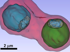
Focusing into a selected plane is achieved by concentrating the acoustic waves coming from the object with a concave cylindrical acoustical mirror onto an optical detector, which is a light beam in an optical interferometer. For the US image, part of an infrared laser pulse is used to excite an acoustic pulse by directing it onto an optically absorbing layer on the acoustic mirror surface. Another part of the same laser pulse is frequency/wavelength converted by nonlinear optical processes and used for PA excitation. Co-registered PA and US images are obtained after applying the inverse Radon transform to the data, which are gathered while rotating the object relative to the detector. Phantom experiments demonstrated a resolution of 1.1 mm between the sections of both imaging modalities and an inplane resolution of about 60 mm and 120 mm for the US and PA modes, respectively. The complementary contrast mechanisms of the two modalities could be shown by images of a zebrafish.
Generation of focused ultrasound pulses with high bandwidth and detection of acoustical signals from a selected slice are made possible by a single element, a concave cylindrical lens coated with a light absorbing metal layer. This makes the device a cost-effective alternative to large scale array tomographs in cases where the region of interest can be covered by a low number of section images and where an imaging time of several seconds per slice is tolerable.
Most read news
Original publication
Organizations
Other news from the department science

Get the analytics and lab tech industry in your inbox
From now on, don't miss a thing: Our newsletter for analytics and lab technology brings you up to date every Tuesday. The latest industry news, product highlights and innovations - compact and easy to understand in your inbox. Researched by us so you don't have to.



![[Fe]-hydrogenase catalysis visualized using para-hydrogen-enhanced nuclear magnetic resonance spectroscopy](https://img.chemie.de/Portal/News/675fd46b9b54f_sBuG8s4sS.png?tr=w-712,h-534,cm-extract,x-0,y-16:n-xl)
