Automatic tracking of biological particles in cell microscopy images
Heidelberg scientists develop powerful automatic method for image analysis
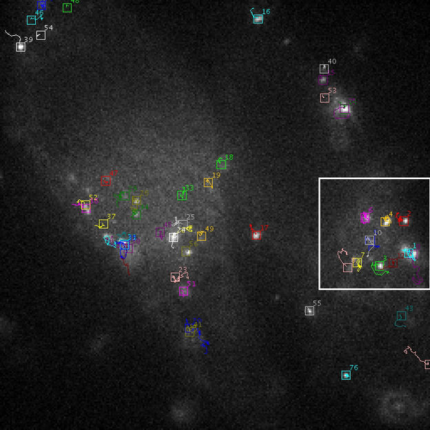
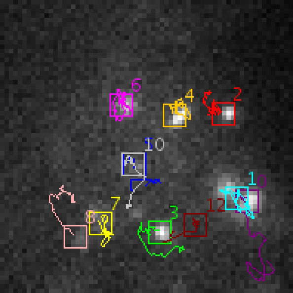
In order to track the movements of biological particles in a cell, scientists at Heidelberg University and the German cancer Research Center have developed a powerful analysis method for live cell microscopy images. This so-called probabilistic particle tracking method is automatic, computer-based and can be used for time-resolved two- and three-dimensional microscopy image data. The Heidelberg method achieved the best overall result in an international competition that compared different methods for image analysis. The competition results were recently published in the journal “Nature Methods”.

Tracking result for virus particles
Ruprecht-Karls-Universität Heidelberg

Tracking result for virus particles – an enlarged section
Ruprecht-Karls-Universität Heidelberg


The task of how to automatically track the movement of biological particles such as viruses, cell vesicles or cell receptors is of key importance in biomedical applications for the quantitative analysis of intracellular dynamic processes. Manually analysing time-resolved microscopy images with hundreds or thousands of moving objects is not feasible. In recent years, therefore, there has been increasing emphasis on the development of automatic image analysis methods for particle tracking. These methods are computer-based and determine the positions of particles over time. To objectively compare the performance of these methods, an international competition was organised in 2012 for the first time.
A total of 14 research teams participated in the “Particle Tracking Challenge”, including Dr. William J. Godinez and Associate Professor Dr. Karl Rohr from Heidelberg University and the German Cancer Research Center (DKFZ). In the competition, the different image analysis methods were applied to a broad spectrum of two- and three-dimensional image data and their performance was quantified using different measures. The three best methods were determined for each category of data. With a total of 150 “Top 3 Rankings”, the Heidelberg scientists achieved the best overall result.
The particle tracking method developed by Dr. Godinez and Dr. Rohr is based on a mathematically sound method from probability theory that takes into account uncertainties in the image data, e.g. due to noise, and exploits knowledge of the application domain. “Compared to deterministic methods, our probabilistic approach achieves high accuracy, especially for complicated image data with a large number of objects, high object density and a high level of noise,” says Dr. Rohr. The method enables determining the movement paths of objects and quantifies relevant parameters such as speed, path length, motion type or object size. In addition, important dynamic events such as virus-cell fusions are detected automatically.
Original publication
Most read news
Other news from the department science

Get the analytics and lab tech industry in your inbox
By submitting this form you agree that LUMITOS AG will send you the newsletter(s) selected above by email. Your data will not be passed on to third parties. Your data will be stored and processed in accordance with our data protection regulations. LUMITOS may contact you by email for the purpose of advertising or market and opinion surveys. You can revoke your consent at any time without giving reasons to LUMITOS AG, Ernst-Augustin-Str. 2, 12489 Berlin, Germany or by e-mail at revoke@lumitos.com with effect for the future. In addition, each email contains a link to unsubscribe from the corresponding newsletter.


