Scientists identify how repair protein finds DNA damage
Researchers at the University of Pittsburgh School of Medicine and University of Pittsburgh Cancer Institute (UPCI) have demonstrated how Rad4, a protein involved in DNA repair, scans the DNA in a unique pattern of movement called "constrained motion" to efficiently find structural faults in DNA. The findings could lead to therapies that boost existing drug treatments and counter drug-resistance.
"Rad4 is like the cop who is the first responder at an accident," said senior author Bennett Van Houten, Ph.D., Richard M. Cyert Professor of Molecular Oncology, Pitt School of Medicine, and co-leader of UPCI's Molecular and Cellular Cancer Biology Program. "The cop can move quickly to recognize where the incident is, and regulate traffic while directing the paramedics arriving in an ambulance."
Constrained motion allows Rad4 to be fast enough to scan large lengths of DNA quickly yet slow enough that it does not miss structural errors in DNA that could be caused by chemicals or ultraviolet (UV) light.
Mutations in Rad4, called XPC in humans, and other proteins in the DNA repair machinery are known to cause a genetic condition called xeroderma pigmentosum, where individuals have sensitivity to sunlight and are at an extremely high risk for developing skin cancer.
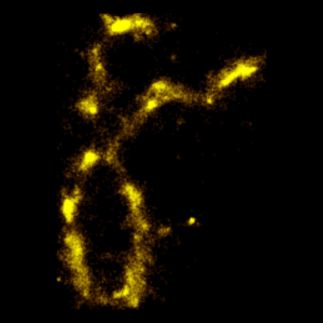
Muwen Kong, a graduate student in Dr. Van Houten's laboratory, along with his collaborators, tagged normal and mutant Rad4 molecules with light-emitting quantum dots. They then watched them move across strands of DNA suspended between beads using a fluorescence microscope.
The results obtained suggest that the first responder, consisting of Rad4 and another protein, Rad23, quickly scans the DNA for accidents by attempting to bend it. Alterations in the structure of DNA, such as those caused by chemicals or UV light, change the ease with which DNA can be bent. Once a potential accident is recognized, the Rad4-Rad23 first-responder team slows down to a 'constrained motion' pattern to more carefully examine a smaller region of 500-1,000 base pairs in the DNA.
When structural damage is confirmed, Rad4-Rad23 stays near the scene and flags down the "paramedics," comprised of the rest of the DNA repair machinery, to fix the damage. This mechanism, which Dr. Van Houten calls 'recognition-at-a-distance,' allows Rad4 to be near the error without impeding the rest of the DNA repair crew.
Though much work is needed before these results can be translated to the clinic, the results provide new avenues to improve treatment methods, especially in cancer. Resistance is a major problem with current treatments, such as the drug cisplatin, which kills cancer cells by introducing DNA crosslinks similar to UV light. By developing drugs that target Rad4/XPC or other repair proteins, it could be possible to enhance the effects of current treatments when they are used together, and also reduce the chances of tumor cells developing resistance, Dr. Van Houten said.
Original publication
Muwen Kong, Lili Liu, Xuejing Chen, Katherine I. Driscoll, Peng Mao, Stefanie Böhm, Neil M. Kad, Simon C. Watkins, Kara A. Bernstein, John J. Wyrick, Jung-Hyun Min, Bennett Van Houten; "Single-Molecule Imaging Reveals that Rad4 Employs a Dynamic DNA Damage Recognition Process"; Molecular Cell; 2016
Other news from the department science
Most read news
More news from our other portals
See the theme worlds for related content
Topic world Fluorescence microscopy
Fluorescence microscopy has revolutionized life sciences, biotechnology and pharmaceuticals. With its ability to visualize specific molecules and structures in cells and tissues through fluorescent markers, it offers unique insights at the molecular and cellular level. With its high sensitivity and resolution, fluorescence microscopy facilitates the understanding of complex biological processes and drives innovation in therapy and diagnostics.
