alpha300 R
3D Raman microscopes with unequalled speed, sensitivity and resolution
WITec Wissenschaftliche Instrumente und Technologie GmbH
Visualize and characterize every chemical detail
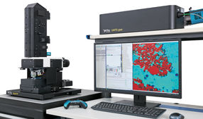
The WITec alpha300 is a unique confocal Raman microscope capable of routinely performing 3D chemical Raman imaging while maintaining the highest measurement speed and spectral quality. This is particularly beneficial for applications in which the exact spatial representation of the chemical components on the surface or within the sample is important. Depth profiles, 3D image stacks or topographic Raman images can be easily created with maximum informational content. The local resolution is always dependent on the physical diffraction limit and has an approximate minimum of 200 nm. At the same time, high-speed measurements are possible in which up to 1300 spectra per second can be recorded.
With the most recent release of a new generation of the alpha300 apyron microscope, WITec takes Raman imaging automation to the next level. Within the alpha300 series, the “apyron” is the top of the line Raman imaging system. It combines ease-of-use and ultimate capability by automating hardware control and offering pre-configured measurement routines. This streamlines the experimental workflow and yields reproducible results with unrivaled speed, sensitivity and resolution.
A critical advantage of WITec Raman technology is the flexible, high-throughput UHTS Spectrometer Series. Each spectrometer is optimized for the excitation wavelength used. As a result the user achieves a much higher throughput than with conventional spectrometers and can operate, for example, with a lower laser power, which is of particular benefit when investigating sensitive samples. The spectral properties are characterized by a peak shape that corresponds to an almost ideal Lorenz curve and nearly the entire relevant spectral range can be recorded with a pixel resolution of up to 0.1 cm-1 / pixel.
If the laser power is to be accurately measured and adjusted, the customer can be provided with the TruePower option, which can be used to determine the absolute laser power in increments of 0.1 mW. The values are recorded with the Raman data acquisition and can be retrieved later – important for internal documentation. The experiment can, of course, be repeated at a later time under precisely the same conditions. This renders obsolete neutral density filters, which are often used for this purpose despite only being able to provide a relative attenuation.
The modular design of the microscope allows Raman imaging to be combined with other microscopy technologies. WITec is a pioneer in this field and has long been able to offer its customers Raman microscopy integrated with AFM or SNOM, selectable through simply rotating the microscope turret. A combination with optical profilometry in WITec’s patented TrueSurface microscopy option for topographic Raman imaging allows large, irregular, rough or inclined samples to be investigated and eliminates the need for complex sample preparation. With RISE microscopy, even correlative Raman-SEM images are possible.
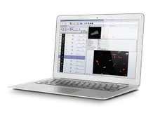
The WITec alpha300 Raman imaging system offers several advantages for versatile research and development applications, without compromise in confocality while maintaining the highest spectral performance and speed and providing detailed 3D analyses of different samples. Straightforward operation is ensured by new software features in the established WITec SUITE 4 and other new innovative operating concepts. Raman imaging is therefore a flexible and versatile tool for chemical imaging and analysis.
Applications
- Pharmaceutical research (drug distribution, drug delivery systems, ...)
- Life Sciences and Biomedicine (live cell examinations, tissues, ...)
- Forensics (material analyses, inks, ...)
- Materials science & nanotechnology (2D materials, nanoparticles, surface analyses, ...)
- Coatings and thin films (layer build-up and thickness, depth profiles, homogeneity ...)
- Photovoltaics and semiconductors (strain analyses, coatings, ...)
- Characterization of polymers (surface structures, crystallinity, ...)

1
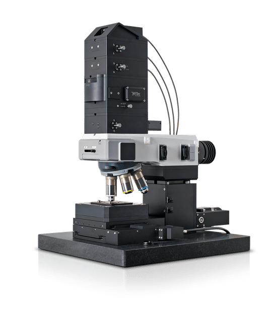
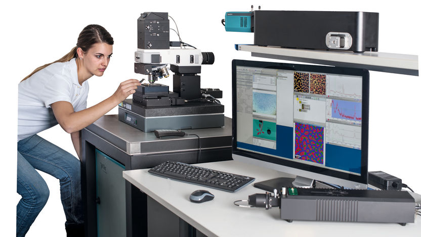
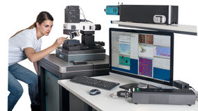
alpha300 Raman microscope for ultrafast, high-resolution, chemical 3D Raman imaging

2
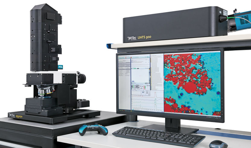
new generation of the alpha300 apyron microscope

3
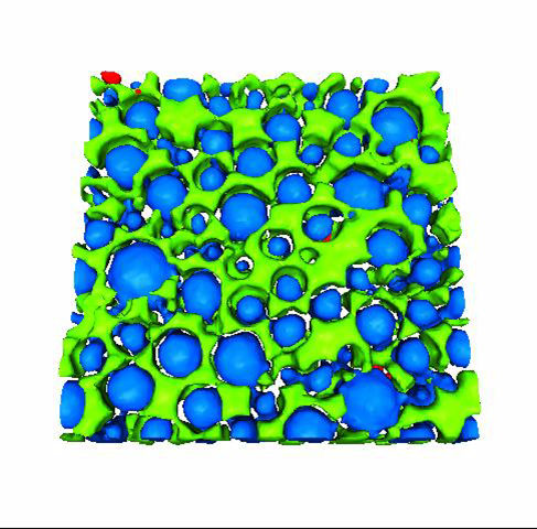
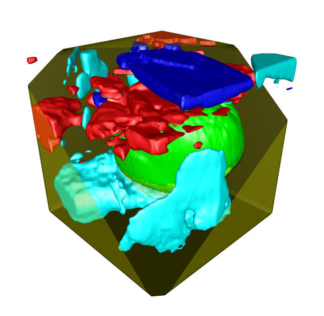
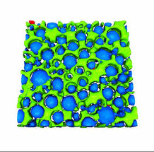
3D Raman image of a pharmaceutical emulsion

4
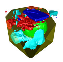
3D Raman image of pollen (green) and sugar crystals in honey

5
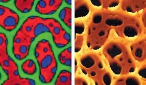
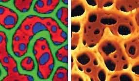
Correlated Raman AFM imaging: Raman (left) and AFM image (right) from the same sample region of a polymer mixture
6
WITec alpha300 Raman microscope: No compromise in confocality combined with the highest spectral performance and speed allows detailed 3D analysis of a wide range of samples
Request information about alpha300 R now

Raman microscopes: alpha300 R
3D Raman microscopes with unequalled speed, sensitivity and resolution
