Keeping a cell’s powerhouse in shape
A German-Swiss team around Professor Oliver Daumke from the MDC has investigated how a protein of the dynamin family deforms the inner mitochondrial membrane.
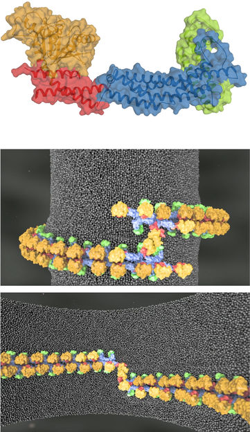
Mitochondria are not rigid organelles – they continuously divide and fuse with each other. A key player of this process is Mgm1, a protein of the dynamin family. Using the technique of X-ray crystallography, the team was able to determine the protein’s three-dimensional structure: the motor (the GTPase domain, orange), the lever (the BSE domain, red), the stalk (blue) and the newly discovered paddle (green). Combining these results with cryo-EM data, yeast growth and fluorescence microscopy data from cooperation partners, we were able to propose molecular models demonstrating how Mgm1 filaments stabilise and remodel the inner membrane of mitochondria.
Daumke Lab, MDC
Mitochondria are the powerhouses of our cells, generating energy in the form of chemical compounds such as ATP. To do this job effectively, mitochondria have a characteristic organisation: In addition to an outer membrane, they have an inner membrane with many invaginations. On this inner membrane are all the enzymes essential to the production of ATP.
Mitochondria are not rigid organelles
“We were interested in how mitochondria acquire and maintain their characteristic shape,” says Professor Oliver Daumke, leader of the MDC research group Structural Biology of Membrane-Associated Processes and one of the two senior authors of the study published in Nature. “The fact is that mitochondria are not rigid organelles – they are continuously dividing and fusing with each other.” One result of this process is that damaged sections are removed.
“But the mitochondria don’t always function so perfectly,” explains Dr. Katja Faelber from the Crystallography Department at the MDC, one of the study’s two first authors. “Many neurodegenerative diseases, such as Alzheimer’s and Parkinson’s disease, are the result of an imbalance between mitochondrial fission and fusion, which causes neurons to slowly die off.”
A molecular machine to reorganize the mitochondrial membrane
Key players in the remodelling of the mitochondrial membrane are the mitochondrial genome maintenance protein 1 (Mgm1) in yeats and the optic atrophy protein 1 (OPA1) in humans. Mutations in the gene for OPA1 lead to a hereditary disease of the optic nerve known as optic atrophy, one of the most common causes of congenital blindness.
“Mgm1 and OPA1 are located on the inner mitochondrial membrane, particularly close to the characteristic invaginations,” explains Oliver Daumke. Here the proteins function as molecular machines, converting chemical energy into mechanical energy.
Prior to this study, it was already known that the proteins consist of several sub-units: of the GTPase domain, which functions as the actual motor, of the BSE (bundle signalling element) domain, which acts as a lever, and the stalk. “We were especially interested in the stalk, because Mgm1 assembles via this unit into filament-like structures,” says Oliver Daumke. These filaments are essential for the function of the molecular machine and consequently for the deformation of the membrane. Without them, the machine would not work.
Using different techniques to build a molecular model
To understand in more detail how Mgm1 stabilises the invaginations and controls the continuous remodelling of the inner mitochondrial membranes, the scientists at the MDC determined the structure of the protein via X-ray crystallography. “Using this method, we generated a three-dimensional atomic model of the Mgm1 protein,” explains Katja Faelber.
The structural biology team in Berlin then cooperated with the group of Professor Werner Kühlbrandt at the Max Planck Institute of Biophysics in Frankfurt am Main, who examined the protein using cryo-electron microscopy. “This technique results in lower resolution structures, but in contrast to X-ray crystallography it enabled us to study Mgm1 in its membrane-bound form,” Katja Faelber says.
This led to the discovery of a fourth, previously unknown domain of Mgm1, which they named the ‘paddle’. “Via this elongated domain Mgm1 attaches itself to the inner mitochondrial membrane,” explains Oliver Daumke. “Additionally, we also identified the contact sites in the stalk that are essential for the arrangement of Mgm1 into filaments.” This gave the team all the puzzle pieces they needed to computer-simulate the processes occurring on the membrane.
Mutations in Mgm1 lead to a collapse of the mitochondria
To verify whether the identified contact surfaces were critical for the stabilisation of the inner mitochondrial membrane, the group led by Professor Martin van der Laan at Saarland University Medical Center in Homburg replaced certain amino acids in these regions. “Indeed, the protein lost its function,” says Oliver Daumke. “The mitochondria did no longer form the characteristic invaginations of the inner membrane and did not fuse with each other.” Many small mitochondrial fragments were left behind, he adds.
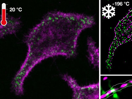
Finally, the group of Professor Aurel Roux at the University of Geneva observed in real time how Mgm1 attaches to membranes using fluorescence microscopy. “The surprising new observation was that the protein binds not only to the outside of artificial membrane tubes, but also to the inside,” says Katja Faelber. “This has never been described for any other member of the dynamin superfamily so far. In fact, this geometry corresponds to that of the invaginations of the inner membrane.”
Improving our understanding of genetic blindness
The team is optimistic that the new findings will benefit medical research. “The results of our study can explain which processes are impaired throughout the course of optic atrophy disease in the eye – how mutations in the OPA1 gene cause the dysfunction of the protein,” says Oliver Daumke. He adds that perhaps one day, this knowledge will make it possible to find a therapy.
Original publication
Other news from the department science
Most read news
More news from our other portals
See the theme worlds for related content
Topic world Fluorescence microscopy
Fluorescence microscopy has revolutionized life sciences, biotechnology and pharmaceuticals. With its ability to visualize specific molecules and structures in cells and tissues through fluorescent markers, it offers unique insights at the molecular and cellular level. With its high sensitivity and resolution, fluorescence microscopy facilitates the understanding of complex biological processes and drives innovation in therapy and diagnostics.
