Cancer DNA in the blood: an early warning system for recurrent tumors in children
Cell-free tumor DNA can be detected early in liquid biopsies
Neuroblastomas release DNA fragments into the blood and cerebrospinal fluid of patients. These genetic material can indicate a possible disease recurrence at an early stage. It also provides information about changes in certain cancer-driving genes. This is what scientists from the German Consortium for Translational Cancer Research (DKTK), a partner site at Charité Berlin, have discovered with the help of state-of-the-art molecular biological methods. They believe that this principle can be used in clinical practice as a gentle method for the early detection of recurrent tumors.

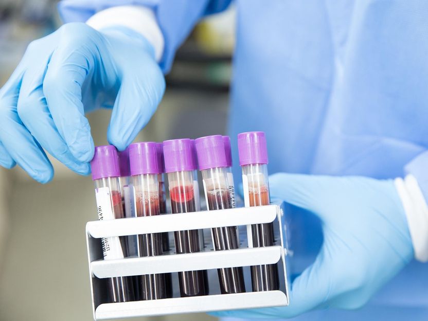
Symbolic image
pixabay.com
In the DKTK, the German Cancer Research Center (DKFZ) in Heidelberg, as the core center, joins forces in the long term with university partner sites in Germany that have a special reputation for oncology.
Neuroblastomas are malignant tumors that occur predominantly in infants and young children. They arise from cells of the embryonic nervous system and usually form in the spinal chord or the adrenal gland. The course of the disease varies widely. About half of neuroblastomas are particularly aggressive and may recur after therapy. In this case, the patients' chances of survival are very low.
Recurrence is triggered by a small number of tumor cells surviving the therapy. Detecting this so-called minimal residual disease at an early stage - if possible before it can lead to relapses - can improve the further prognosis of the affected children. A prerequisite for this is continuous monitoring with diagnostic methods, because several surgical procedures to remove tissue in series would be far too stressful for those affected. "A promising alternative to tissue analysis is provided by so-called liquid biopsies, which are obtained in a minimally invasive way, for example by taking a sample of the patient's blood or cerebrospinal fluid," explains pediatrician Hedwig Deubzer from the DKTK partner site at the Charité in Berlin. She and her team are researching such methods.
Scientists had already succeeded in detecting cell-free genetic material from tumor cells, so-called ctDNA, in the blood and cerebrospinal fluid of children with neuroblastoma using molecular biological techniques. The DKTK team now investigated whether ctDNA from liquid biopsies could be used as a diagnostic marker to follow the course of the disease - with success. It was able to show that cell-free tumor DNA can be detected early in liquid biopsies from high-risk neuroblastoma patients. "We even identified two patients who had no signs of disease progression up to that point," Deubzer said. "We anticipate that liquid biopsy will be very suitable for routine clinical use because of its sensitivity and the fact that sample material can be easily obtained."
The researchers were also able to show that ctDNA fragments are not only useful as an early warning system for the return of neuroblastoma after therapy. They also provide information about molecular changes in tumor DNA that occur over time and that may be relevant to therapy. The researchers also found such dynamic changes in the region of two known cancer driver genes: the MYCN gene and the ALK gene. "The ALK gene in particular represents a target for cancer therapy with an ALK inhibitor," Deubzer said. "The early detection of alterations in this gene therefore also means a new option for follow-up treatment for some patients."
Original publication
Marco Lodrini, Josefine Graef, Theresa M. Thole-Kliesch, Kathy Astrahantseff, Annika Sprüssel, Maddalena Grimaldi, Constantin Peitz, Rasmus B. Linke, Jan F. Hollander, Erwin Lankes, Annette Künkele, Lena Oevermann, Georg Schwabe, Joerg Fuchs, Annabell Szymansky, Johannes H. Schulte, Patrick Hundsdoerfer, Cornelia Eckert, Holger Amthauer, Angelika Eggert, Hedwig E. Deubzer; Clin Cancer Res; 2022
Most read news
Original publication
Marco Lodrini, Josefine Graef, Theresa M. Thole-Kliesch, Kathy Astrahantseff, Annika Sprüssel, Maddalena Grimaldi, Constantin Peitz, Rasmus B. Linke, Jan F. Hollander, Erwin Lankes, Annette Künkele, Lena Oevermann, Georg Schwabe, Joerg Fuchs, Annabell Szymansky, Johannes H. Schulte, Patrick Hundsdoerfer, Cornelia Eckert, Holger Amthauer, Angelika Eggert, Hedwig E. Deubzer; Clin Cancer Res; 2022
Organizations
Other news from the department science

Get the analytics and lab tech industry in your inbox
By submitting this form you agree that LUMITOS AG will send you the newsletter(s) selected above by email. Your data will not be passed on to third parties. Your data will be stored and processed in accordance with our data protection regulations. LUMITOS may contact you by email for the purpose of advertising or market and opinion surveys. You can revoke your consent at any time without giving reasons to LUMITOS AG, Ernst-Augustin-Str. 2, 12489 Berlin, Germany or by e-mail at revoke@lumitos.com with effect for the future. In addition, each email contains a link to unsubscribe from the corresponding newsletter.


